
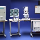
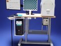
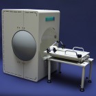
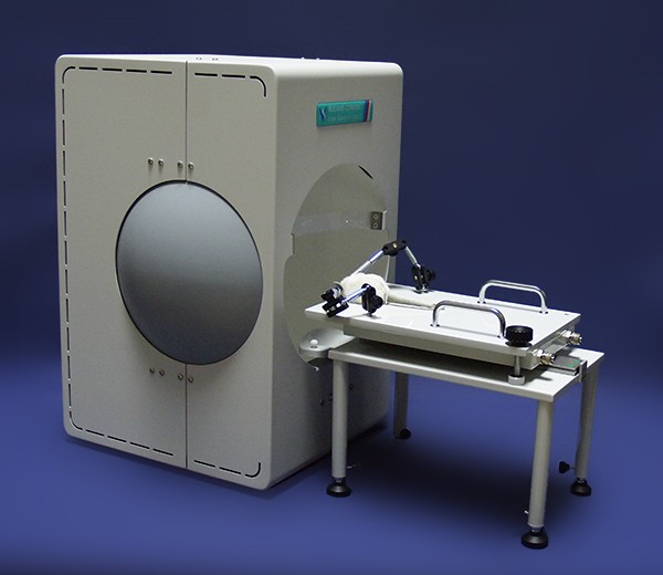
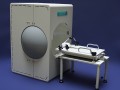
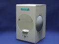
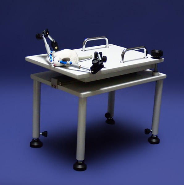
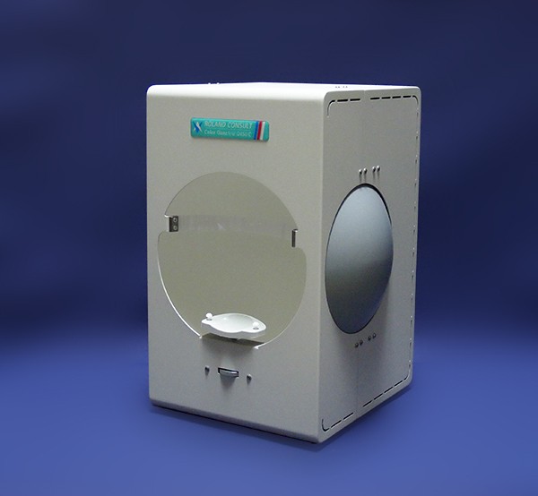
RETI-map - animal
Electrophysiological Unit combined - with Scanning Laser Ophthalmoscope
|
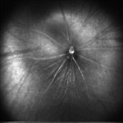
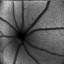
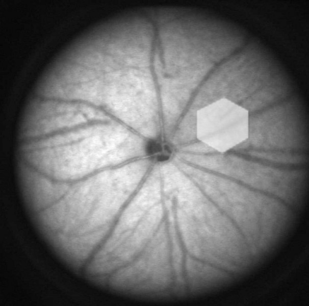
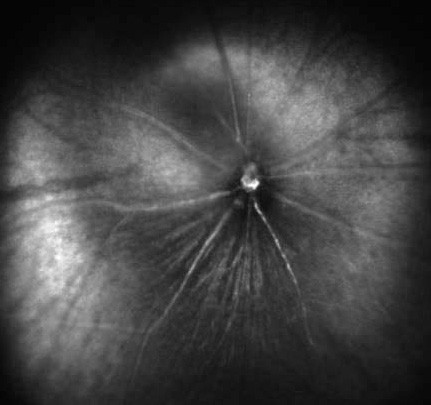
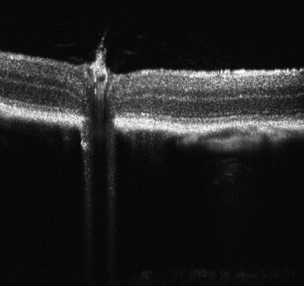
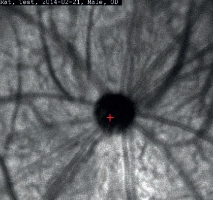
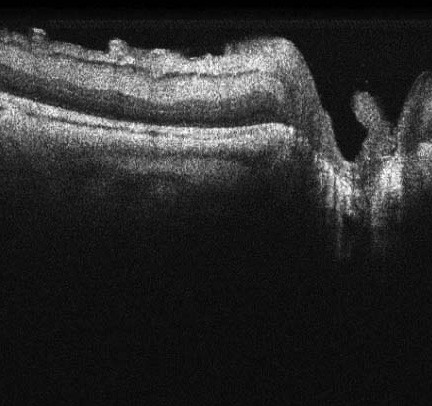
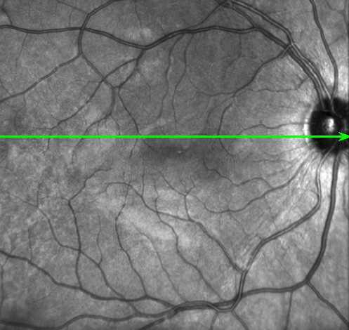
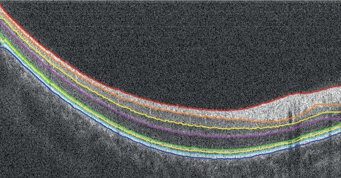
The RETImap animal® infrared imaging with infrared laser enables the user to produce a higher quality fundus image in comparison to alternative white light illumination photo systems.
|
|
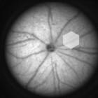


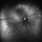
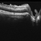
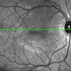
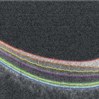

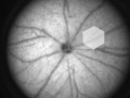
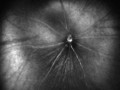
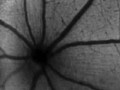
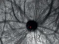
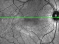
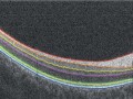

Infrared imaging cSLO
| Field of view: |
35° x 35° square |
| Laser source: | SLD 920 nm (IR Image) |
| Image Resolution: | 512 x 512 |
| Digital image: | 12 bit |
| Optical resolution: | ca. 30 µm |
| Pupil size requirement: |
>= 2mm |

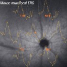
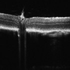
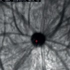
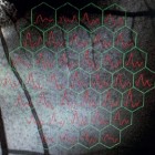
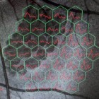
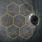
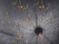
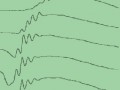
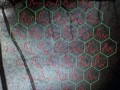
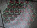
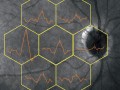
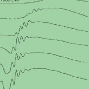
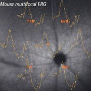
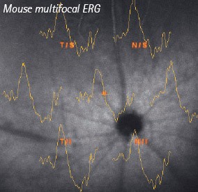
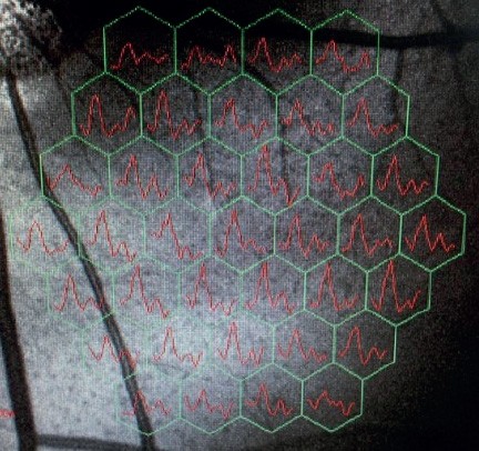
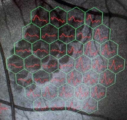
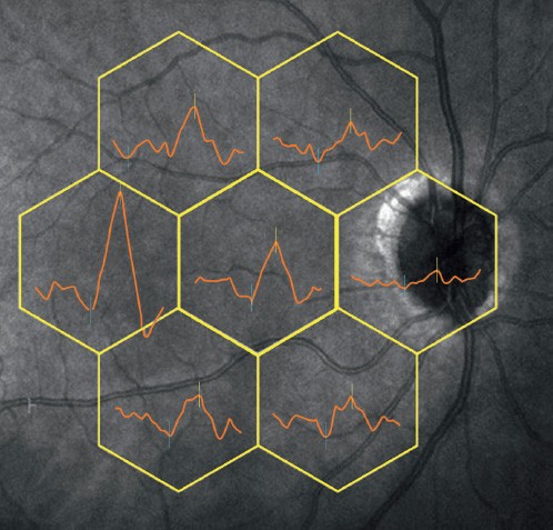
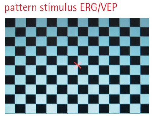
 The combination cSLO + ERG/VEP is a worldwide unique technology.It allows simultaneous infrared laser monitoring during electrophysiological diagnostic. With the built-in stimulator, the high resolution DLP projector, it is possible to stimulate a large number of retinal locations and extract their responses. The final result is a function map superimposed onto the fundus cSLO image. Regionally confined areas of dysfunction can be detected.
The Roland Consult Fundus EyeTracker (RCFET) software helps to detect all eye movement artefacts during the test. The various electrophysiological functions are:
- multifocal ERG,
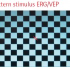
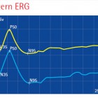
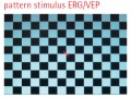
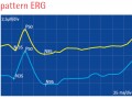
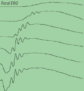
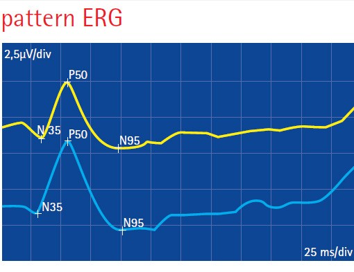
- focal flash ERG, focal pattern ERG,
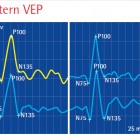
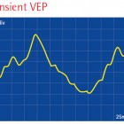
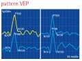
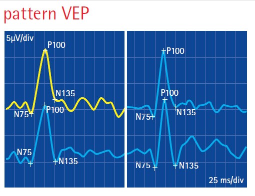
- Pattern VEP,
- focal Pattern VEP and focal Flash VEP.
This device can also be upgraded with the classical electrophysiological stimulators and programs.

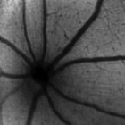
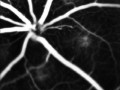
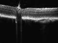
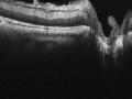
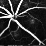
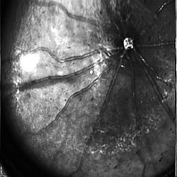
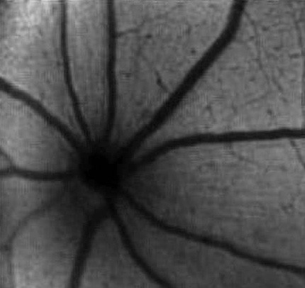
 Module Angiography - The Angiography Module runs with a blue laser source (488 nm). It generates higher quality fundus images in comparison to alternative white light illumination photo systems and since no flashes are involved, the system is more patient friendly.
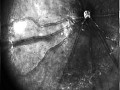
At the beginning of an injection the laser source automatically switch to Angiography mode and a video will be recorded with an exact timestamp on every image. This helps the user to concentrate more on the patient during the examination and review the angiography images later. The advantage of the laser system is also the high speed of image acquisition. Instead of watching static images from early, mid or late phase it is easy to observe the dye in the
vessels to localize narrowings and partial blockades.
| Laser source: | Blue: 488 nm |
| Digital image: | 512x512 |
| Record Mode: | 15 pictures / sec |
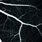

Is a protein composed of 238 amino acid residues (26.9 kDa) that exhibits bright green fluorescence when exposed to light in the blue to ultraviolet range.
Laser source: 488 nm
- Superposition of the reflected images, real time averaging
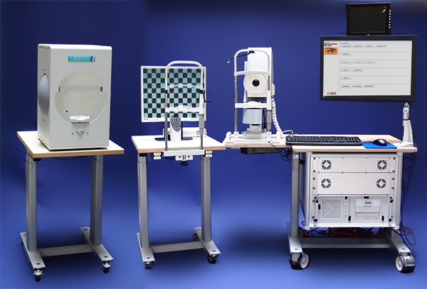
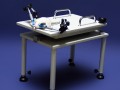
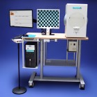
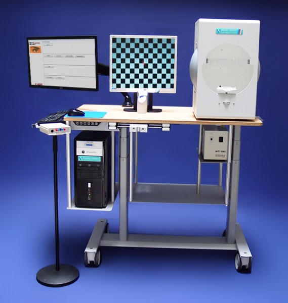
RETI-port/scan 21
Electrophysiological Test Unit
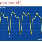
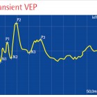
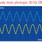
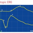
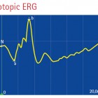
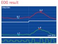
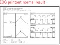
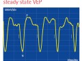
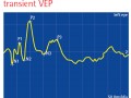
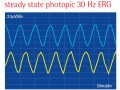
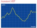
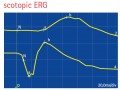
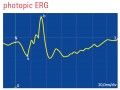
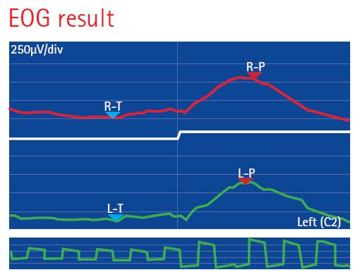
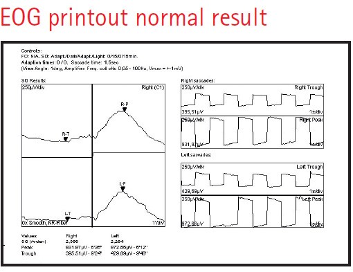
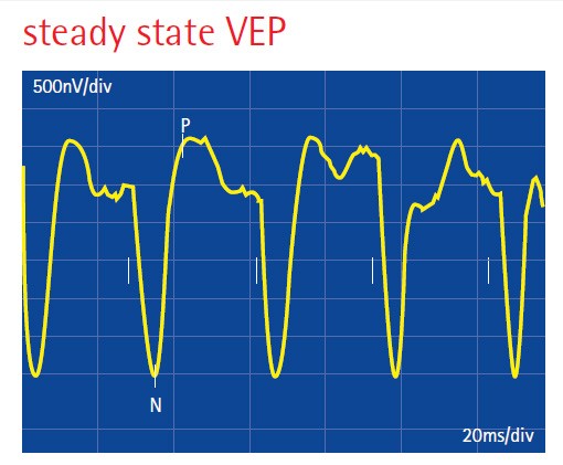
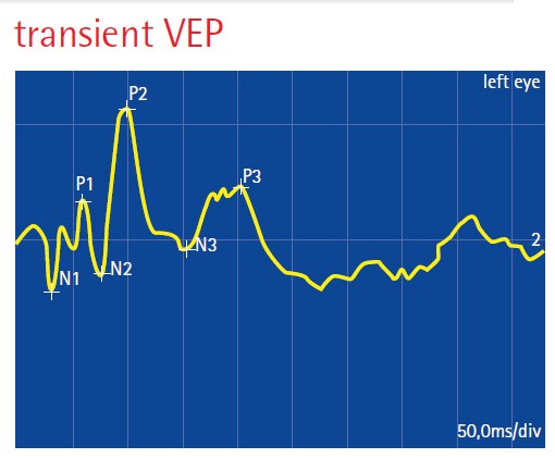
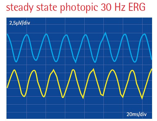
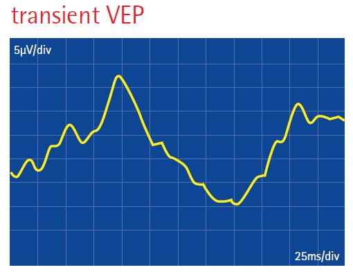
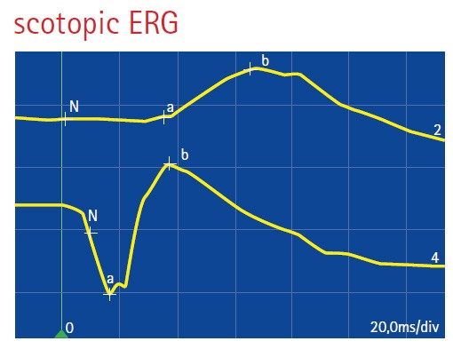
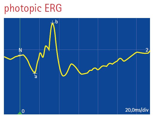
Pattern VEP + Pattern ERG + Flash VEP, ISCEV ERG, EOG Fast and Slow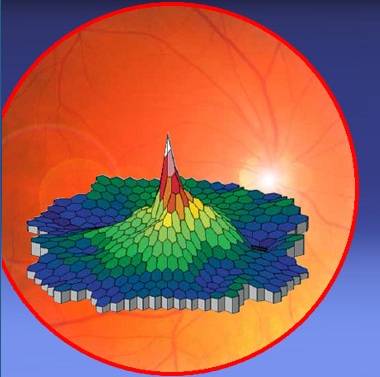 Electrophysiological Test Unit is usable for Pattern VEP + Pattern ERG + Flash VEP, for scotopic and photopic ERG, EOG fast and slow, mf ERG Flash stimulation and mf VEP Pattern stimulation. All ISCEV standards and guidelines are included.
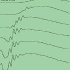
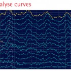
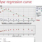
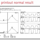
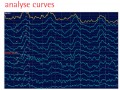
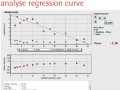
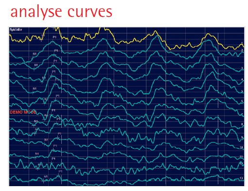
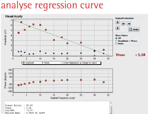
The biosignal amplifier includes a preamplifier near the patient. All patient data and the results are stored in a database. The biosignal and averaged curves from all channels can be displayed on the monitor. In the analyze mode the system automatically sets all markers and calculates all the defined parameters.
The RETI-port/scan 21 unit from the Roland Consult Stasche & Finger GmbH includes the stimulator units and the data recording and analyzing system.
| Modell: | basic | alpha | alpha plus | beta | beta plus | gamma | gamma plus | gamma plus² | delta plus | delta plus² |
| No. | 0 | 1 | 2 | 3 | 4 | 5 | 6 | 7 | 8 | 9 |
| Protocols | ||||||||||
| Pattern-VEP | x | x | x | x | x | x | x | x | o | o |
| Pattern-ERG | x | x | x | x | x | x | x | x | o | o |
| Flash-VEP | o | x | x | x | x | x | x | x | o | o |
| Albino VEP 1 chanel | o | o | o | o | o | x | x | x | o | o |
| Flash ERG 1 channel | o | x | x | - | - | - | - | - | - | - |
| Flash ERG 2 channel | - | - | - | x | x | x | x | x | - | - |
| Photopic Negative Resp. | - | - | - | x | x | x | x | x | - | - |
| ON-OFF Resp. | - | - | - | o | o | x | x | x | - | - |
| S-Cone ERG | - | - | - | o | o | x | x | x | - | - |
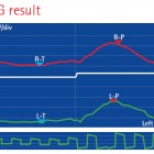
| EOG | - | - | - | x | x | x | x | x | - | - |
| Multifocal ERG P | o | - | x | - | x | - | - | - | x | - |
| Multifocal ERG S | - | - | - | - | - | - | x | x | o | x |
| Multifocal VEP | o | - | - | - | - | - | - | x | o | x |
| Multifocal Science | o | - | - | - | - | - | x | x | o | x |
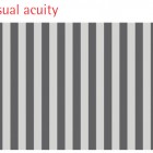
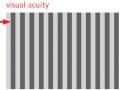
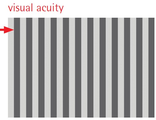
| Visual Acuity | o | o | o | o | o | x | x | x | o | o |
| Glaucoma Screening | - | o | o | o | o | x | x | x | - | - |
| Contrast Sensitivity | o | o | o | o | o | o | o | x | o | o |
| Nystagmograohy | o | o | o | o | o | o | o | x | o | o |
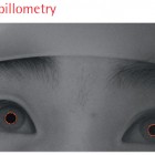
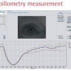
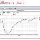
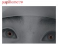
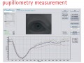
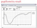
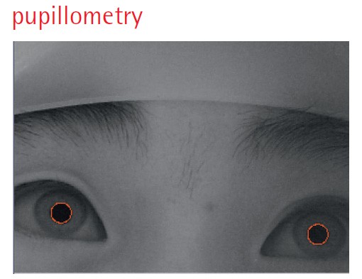
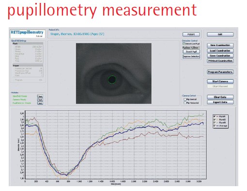
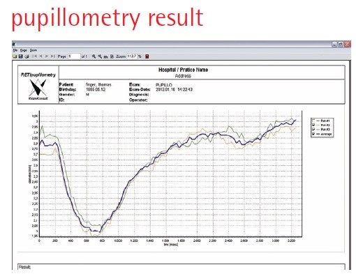
| Pupillometry | - | - | - | o | o | o | o | x | - | - |
| Scientific Tool Port | - | - | - | - | - | x | x | x | - | - |
| Scientific Tool Scan | - | - | - | - | - | - | x | x | - | x |
| Stimulators | ||||||||||
| Monitor | x | x | x | x | x | x | x | x | x | x |
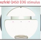
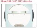
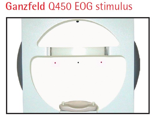
| Ganzfeld Q450 C | - | - | - | x | x | - | - | - | - | - |
| Ganzfeld Q450 SC | - | - | - | - | - | x | x | x | - | - |
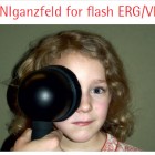
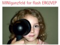
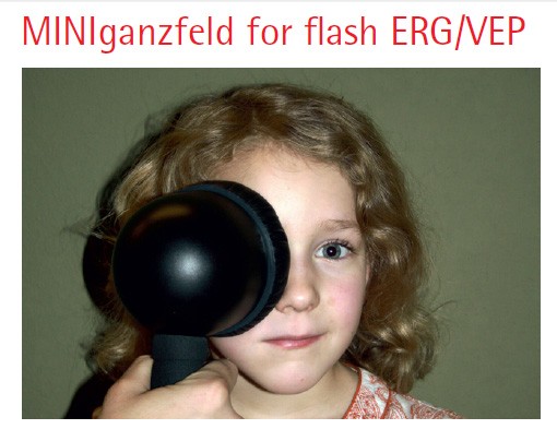
| MINIganzfeld I8 | - | x | x | o | o | o | o | o | - | - |
| BABYflash E130 | - | o | o | o | o | o | o | o | - | - |
| Amplifier | ||||||||||
| 2 Channels | x | x | x | x | x | - | - | - | x | - |
| 4 Channels | o | o | o | o | o | x | x | x | o | x |
| x Standard | ||||||||||
| o Option | ||||||||||
| - N.A. |
| Diagnosis | EOG | ERG | Bright Flash ERG | Pattern ERG | Flash VEP | Pattern VEP | Special VEP | mf VEP |
| Inherited retinal dystroohies | + | + | + | + | ||||
| Vascular diseases including diabetes | + | + | + | |||||
| Opaque media or trauma | + | + | + | |||||
| Retrobulbar neuritis | + | + | + | |||||
| Unexplained visual loss | + | + | + | + | ||||
| Infant with questionable vision | + | + | + | + | + | |||
| Albinism | + | + | ||||||
| Toxic and nutritional eye disease | + | + | + | + | + | |||
| Glaucoma | + | + | ||||||
| Suspected intracranial lesion | + | + | + | |||||
|
|
|
|
|
|
|
|
|
|
|
|
|
|
|
|
|
|
|
|
|
|
|
|
|
|
|
|
|
|
|
|
|
|
|
|
|
|
|
|
